BLACK LUNG PICTURE

Black Lung Picture


Black lung disease is the common name for coal workers' pneumoconiosis or anthracosis, a lung disease of older workers in the coal industry, caused by inhalation,
over many years, of small amounts of coal dust. As seen in the picture is
turns the lungs black instead of their normal pink color.


black_lung.bmp


-

BEHCET'S DISEASE PICTURE AND INFORMATION


Behcet's Disease
Pictures


Behcet's disease is a rare,
chronic, lifelong disorder that involves inflammation of blood vessels throughout the
body. Symptoms of Behcet's disease include recurrent oral ulcers
(resembling canker sores), recurrent genital ulcers, and eye
inflammation. The disorder may also cause various types of skin lesions,
arthritis, bowel inflammation, and meningitis (inflammation of the
membranes of the brain and spinal cord)


-

BAKER'S CYST PICTURE


Baker's Cyst Picture


Baker s cysts are benign cystic tumors found
on the back of the knee joint. The cyst forms with an accumulation of fluid
between the 2 groups of muscles behind the knee. This cyst may form from an
injury or inflammation of the membrane lining of the knee joint


Baker's Cyst


-

BELL'S PALSY PICTURES


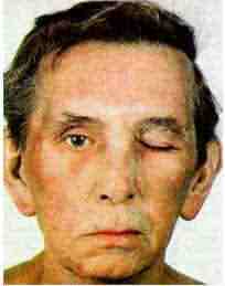
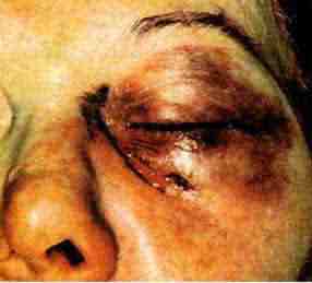
Bell's Palsy Pictures


Bells palsy is a form of facial paralysis resulting from damage to the 7th
(facial) cranial nerve. This nerve disorder afflicts approximately 40,000 Americans each
year. It can strike almost anyone at any age; however, it disproportionately attacks
pregnant women and people who have diabetes, influenza, a cold, or some other upper
respiratory ailment. In addition to one-sided facial paralysis with possible inability to
close the eye, symptoms of Bells palsy may include pain, tearing, drooling,
hypersensitivity to sound in the affected ear, and impairment of taste. The common cold
sore virus, herpes simplex, and other herpes viruses are the likely cause of many cases of
Bells palsy.
Bell's Palsy

-

BENIGN JUVENILE MELANOMA PICTURE


Benign Juvenile Melanoma Picture


A benign, slightly pigmented or red superficial small skin tumor composed of
spindle-shaped, epithelioid, and multinucleated cells that may appear atypical; most
common in children, but also appearing in adults.


Benign Juvenile Melanoma

As seen in this Benign Juvenile Melanoma Picture the small skin tumor is
raised and dark colored.


-

BERLOQUE DERMATITIS PICTURE


Berloque Dermatitis Picture


Berloque dermatitis, a type of photosensitization resulting in deep brown pigmentation
on exposure to sunlight after application of bergamot oil and other essential oils in
perfume.
Berloque dermatitis

Berloque dermatitis


-

BIDACTYLY PICTURE


Bidactyly Picture


Abnormality in which the medial digits are lacking, with only the first and fifth
represented.


Also know as lobster claw deformity
Bidactyly

Bidactyly


-

BLACK EYE PICTURE


Black Eye Picture


Ecchymosis, or more commonly known as a black eye, usually
occurs from some type of trauma to the eye, causing the tissue around the eye to become
bruised. Your child's physician will examine the eye closely to make sure there is no
damage to the actual eye itself.

Black Eye

Black Eye Picture


-

BLEPHAROSPASM PICTURE

Blepharospasm Picture


Blepharospasm is a condition characterized by uncontrollable, forcible closure of the
eyelids caused by a progressive dysfunction of the nerve that controls muscles around the
eye. It is more than just blinking often.


Blepharospasm.jpg (1860 bytes)
-

BLEPHARITIS PICTURE

Blepharitis Picture


Blepharitis is an inflammation of the eyelids, due to infection, usually accompanied by
crusts or scales on the eyelid margin. You can see the inflammation on the
edges of the eyelids called blepharitis in the picture.


Blepharitis.jpg
-

BASAL CELL PUCTURE

Basal Cell Picture


A small, round cell found in the lower part (or base) of the epidermis, the outer layer
of the skin.


Basal cell carcinomas are most common
type of skin cancer, a disease in which the cancer cells resemble the basal cells of the
epidermis, the outer layer of the skin

The picture of basal cells is a
little hard to understand so we linked it to the website it comes from where you
can get an explanation of the picture.
Basal-cell.gif (37352 bytes)


Click on the image above to see the full sized picture

-

BONE MARROW PICTURE

Bone Marrow Picture


Bone Marrow is the inner, spongy substance in the center of the bone that produces all
of the red blood cells and platelets and most of the white blood cells.


bone-marrow.gif

-

BONE CELL PICTURE


Bone Cell Picture And Video


There are three special types of cells that are found only in the bone.

These cell names all start with OSTEO because that is the Greek word for bone.


-

BONE SCAN PICTURE

Bone Scan Picture


A bone scan is a nuclear scanning test that identifies new areas of bone growth or
breakdown. It can be done to evaluate damage to the bones, to detect cancer that has
spread (metastasized) to the bones, and to monitor conditions that can affect the bones
(including infection and trauma).


Bone-Scan_small.jpg


Bone Scan Picture


-

BONE CANCER PICTURE

Bone Cancer Picture


-

BALANITIS PICTURE

Balanitis Picture


Balanitis is when the head of the penis is inflamed. Balanitis can
occur at any age, but is more likely to affect boys under four years of
age, as well as adult males who have not been circumcised.


-

BLASTOMYCOSIS PICTURE


Blastomycosis Picture


Blastomycosis is a rare fungal infection caused by inhaling a
fungus (Blastomyces dermatitidis), which is found in wood and soil.
Blastomycosis

Blastomycosis Picture


-

BONE TISSUE PICTURE

Bone tissue Picture


Bone tissue Picture


bone tissue
-

BLOOD BLISTER PICTURE

Blood Blister Picture


A blood blister is a type of blister that forms when
subdermal tissues and blood vessels are damaged without piercing the
skin. It consists of a pool of lymph, blood and other bodily fluids
trapped beneath the skin. If punctured, it suppurates a dark red fluid.


Blood-Blister.jpg

Blood Blister Picture
-

BLOOD CELL PICTURE

Blood Cell Picture


A blood cell is any cell of any type normally found in blood. In
mammals, these fall into three general categories: *red blood cells,
whose main purpose is the transport of oxygen*white blood cells, which
act as immune cells and fight infection.


blood-cell.jpg


Blood Cell Picture

-

BLOOD CIRCULATION PICTURE

Blood Circulation Picture


A circulatory system (sometimes cardiovascular system) is an organ
system that moves substances to and from cells; it can also help
stabilize body temperature and pH (part of homeostasis). There are three
types of circulatory systems (from simplest to most complex): no
circulatory system, open circulatory system, and closed circulatory
system.


Blood-Circulation.gif
-

BLUE SCLERA PICTURE

Blue Sclera Picture


Blue Sclera is a condition where the white part of the eyes turn blue.
The picture shows the where the white part of the eye has turned blue from
blue sclera.


blue-sclera.jpg (62189 bytes)


-

BLOOD CLOTS PICTURE

Blood Clots Picture


Blood clots are a solid
mass of blood. Its function is to develop at the site of wounds to stop
blood loss. However, clots may form inappropriately within the blood
vessels, for example if the blood is flowing too slowly. Blood clots
within blood vessels can block the blood supply to certain parts of the
body.
blood_clot.jpg


Blood Clots


-

BLOOD CLOTS IN LEG PICTURE

Blood clots in leg picture


Deep venous thrombosis (DVT) refers to a blood clot embedded in one
of the major deep veins of the lower legs, thighs, or pelvis. A clot
blocks blood circulation through these veins, which carry blood from the
lower body back to the " WIDTH="387" HEIGHT="450" lowsrc="brain-cells.jpg" longdesc="Brain Cell Picture">
-

BRAIN TUMOR PICTURE


Brain Tumor Picture


A brain tumor is any growth of abnormal cells or uncontrolled proliferation of cells in
the brain. Primary brain tumors involve any such growth that originates in the brain,
rather than spreading to the brain from another part of the body.
Brain Tumor

Brain Tumor Picture


-

BREAST ABSCESS PICTURE


Breast Abscess Picture


Breast abscess, a fairly common form of abscess, occurs in the tissue of the breast;
this is seen most often in post-partum women and in nursing mothers. Most of these
abscesses are due to an invasion of the breast tissue by skin organisms which either enter
via some minute crack in the nipple or work their way down through the breast duct system.
A breast abscess manifests itself as a warm tender swelling, sometimes accompanied by
chills or fever. It may respond to local compresses and antibiotics by mouth. If it does
not, the swelling will continue and may become fluctuant (soft), at which point an
incision to produce drainage may be necessary. Once drainage is established, the abscess
empties itself and healing generally occurs without incident.
Breast Abscess

Breast Abscess Picture


-

CANCER CELL PICTURE

Cancer Cell Picture


Cancer is a class of diseases characterized by uncontrolled cell division and the
ability of these cells to invade other tissues, either by direct growth into adjacent
tissue (invasion) or by migration of cells to distant sites (metastasis). This unregulated
growth is caused by damage to DNA, resulting in mutations to vital genes that control cell
division, among other functions. One or more of these mutations, which can be inherited or
acquired, can lead to uncontrolled cell division and tumor formation. Tumor
(swelling in Latin) refers to any abnormal mass of tissue, but may be either
malignant (cancerous) or benign (noncancerous). Only malignant tumors are capable of
invading other tissues or metastasizing.


.
-

CANCER MOLE PICTURE

Cancer Mole Picture


It is important to become familiar with the moles on your skin because, in rare cases,
they can become cancerous. Monitoring your moles and other pigmented patches, such as
freckles and age or liver spots, is an important first step in reducing your risk of skin
cancer especially malignant melanoma, which may begin in or near a mole or other
dark spot on the skin. Melanoma also can arise in areas of normal pigmentation.


Click on the image above to see the full sized picture,
other pictures and for more information about this Cancer Mole Picture

-

BREAST CELLULITIS PICTURE AND INFORMATION


Breast cellulitis


-

BREAST CYST PICTURE

Breast Cyst Picture


Breast cysts are fluid-filled sacs that develop within the breast tissue,
usually in the upper half of the breast.
Breast cysts are usually associated with breast cancer and other serious
ailments but this is not always the case. The picture below shows a comparison
of normal breast tissue and tissue with cysts.


breast-cyst.gif


-

CANCER SORE PICTURE

Cancer Sore Picture


Most cancer sores appear in the he mouth or on the lips. Smoking is one of
the major causes. 75% of all oral cancer sores are caused by smoking when only
20 percent of all people smoke.


Click on the image above for the full sized picture and other pictures.

-

BRODMANN'S AREAS


Brodmann's areas picture


Area's of the cerebral cortex mapped out on the basis of the cortical cytoarchitectural
patterns.


Brodmann's areas

Brodmann's areas


A Brodmann's area is a region in the brain cortex defined in many different
species based on its cytoarchitecture. Cytoarchitecture is the organization of
the cortex as observed when a tissue is stained for nerve cells.


-

BRONCHIAL TREE PICTURE


Bronchial tree picture


The intricate network of air passages that supply the lungs. When
inverted, this structure resembles a tree, with the trachea
resembling the trunk.
Bronchial tree

Bronchial tree picture


-

PHOTO AND PICTURE OF BROWN RECLUSE SPIDER BITE


Brown recluse spider bite picture


The brown recluse spider lives throughout the Southern half of the United States. There
are ten different species, six of which are confirmed to be poisonous. The spiders are tan
to brown and most species exhibit a violin-shaped marking on the dorsum of the
cephalothorax. They tend to live in dark dry spaces such as attics, closets and wood
piles, and generally they are not aggressive but rather bite when forced to.


The bite generally causes some mild stinging or burning and ulcerative necrosis may
follow within several hours to several weeks. As seen in the picture
below initial lesion may be red, edematous or
blanched and may later may develop a blue-gray halo around the puncture.
Brown recluse spider bite

Photo and picture of Brown recluse spider bite


-

BULLSEYE RASH PICTURE


Bullseye Rash Picture


A bullseye Rash is caused by a tick bite from the Ixodes. It is the first
symptom of lyme disease. The rash expands from the site of the bight forming
a dark red patch then develops a second circular rash around the initial
rash to form the appearance of a bullseye.

bullseye-rash.jpg


-

BRUSHFIELD SPOTS


Brushfield spots picture


Speckled iris. Little white spots that slightly elevated on the surface of the iris
arranged in a ring concentric with the pupil. These spots occur in normal children but are
far more frequent in Down's syndrome . They were described in 1924 by Thomas Brushfield
and are due to aggregation of a normal iris element.
Brushfield spots

Brushfield spots


-

CANAVAN DISEASE PICTURE

Canavan Disease Picture


Canavan disease (C is an inherited disease. It occurs due to a deficiency of
the enzyme called Aspartoacylase, since the gene which usually tells the body to produce
this enzyme is does not work correctly.. Babies are born apparently
unaffected. At 2 to 4 months of age develops poor head control and seizures.


Canavan.bmp


Sub cortical spongy changes of the white
matter. The cortex above is spared. The dark stained neurons are not affected.

-

BURSA PAIN PICTURE

Bursa Pain Picture


Bursa pain is caused by bursitis. Bursa are small sacks of oil that
are located between tendons and bones near joints. Inflammation due to
trauma or other source can cause significant pain especially when body
movement causes a tendon or muscle to move across them.


-

BUNION PICTURE


Bunion Picture


Bunions occur when the angle between the first and second long bones increases to a
point where it is greater than normal. The increased angle may cause the great toe to abut
the second toe. The bump may become red and very sore, depending on your footwear
selection.

Bunion picture

This bunion Picture
shows extreme inflammation to the joint.


-

CARCINOID PICTURE

Carcinoid Picture


A carcinoid is a slow-growing type of tumor usually found in the gastrointestinal system (most often
in the appendix), and sometimes in the lungs or other sites. Carcinoid tumors may spread
to the liver or other sites in the body, and they may secrete substances such as serotonin
or prostaglandins, causing carcinoid syndrome.


Carcinoid Picture


Visit out Cancer Information Site.

Cancer
Symptoms Hundreds
of pages of cancer information


-

CARCINOMA PICTURE

Carcinoma Picture


A type of cancer that develops from tissues covering or lining organs or glands of the
body, such as skin, colon, lung, or breast. Most of the cancers are carcinomas.


Click the links below for example pictures of Carcinoma.
-

BURSITIS KNEE PICTURE

Bursitis Knee Picture


Bursitis is an inflammation of a bursa. A bursa is a fluid
filled sack that pads areas where tendons rub over bone.


In the knee there are several bursae that can cause bursitis of the
knee. . The image below will show you the 4 bursa listed below.


  • suprapatellar bursa

  • pre-patellar bursa

  • infrapatellar bursa

  • deep infrapatellar bursa


knee-bursitis.png Bursitis Knee Picture
-

CARPAL TUNNEL SYNDROME PICTURE

Carpal Tunnel Syndrome Picture


A compression of the median nerve caused by
swelling tendons in the carpal tunnel. Symptoms include numbness, pain, and tingling in
the thumb and middle fingers, weak grip and diminished strength in the hand.


Carpal-Tunnel-Syndrome.gif (6154 bytes)

Carpal Tunnel Syndrome
picture and
information

-

BURSITIS PICTURE


Bursitis Picture


Bursae are fluid-filled cavities near joints where tendons or
muscles pass over bony projections. They assist movement and reduce friction between
moving parts.


Bursitis can be caused by chronic overuse, trauma, rheumatoid arthritis, gout, or
infection. Sometimes the cause cannot be determined. Bursitis commonly occurs in the
shoulder, knee (washmaid's knee), elbow, and hip. Other areas that may be affected include
the Achilles tendon and the foot.


Bursitis

This bursitis picture
shows extreme inflammation to the bursae.


-

BUTT ACNE PICTURE

Butt Acne Picture


We added this page simple because people search for the this term.
Acne can develop anywhere on the body. Severe acne on the butt is not
common but as seen by the picture is possible.


Google advertising will not let us keep this picture

-

BUTTERFLY RASH PICTURE


Butterfly Rash Picture


The butterfly rash of lupus is typically painless and does not itch. Along with
inflammation in other organs, the butterfly rash of lupus can be precipitated or worsened
by exposure to sunlight, a condition called photosensitivity. The photosensitivity can be
accompanied by a worsening of inflammation throughout the body, causing a
flare of the disease.


Butterfly Rash

Butterfly Rash Picture


-

CALCANEAL BURSITIS PICTURE AND INFORMATION


Calcaneal Bursitis Picture


Calcaneal

bursitis
is a type of
bursitis
occurring under the heel bone.


Calcaneal Bursitis


Individuals with this calcaneal bursitis will usually have an inflamed bursa
on the bottom of the heel. This will typically make standing and walking
difficult. Pain is generally the most significant symptom of calcaneal bursitis.


-

CALCANEUS BONE PICTURE


Calcaneus bone picture


The calcaneus bone
forms the lower, outer part of the ankle and extends downward to form the heel. It is
responsible for bearing much of the immediate stress placed upon the foot during walking.
This bone
is also called the calcaneum.


Calcaneus bone

Calcaneus bone


-

CALLUS PICTURE


Callus Picture


Callus occurs due to hyperkeratosis.
Hyperkeratosis is the thickening of the skin due to pressure. Callus can occur over bony
prominence, on the ball of the foot or on the heel. Callus often occur due to prolonged
standing and may result in chronic pain. A callus appears as yellow or dark brown
discolored skin.


Callus Picture


Callus Picture


This callus picture shows the
allow or dark brown
discolored skin caused by the thickening of the skin.


-

CANDIDA PICTURE

Candida Picture


Candida is a natural flora found in the body. When it gets out
of balance and becomes to prominent then most people call it a yeast infection.
Candida is naturally found in the intestines and vagina of women. As seen in the
picture any place that stays damp to long can develop a candida infection.


candida.jpg Candida Picture
-

CANDIDA ALBICANS PICTURE

Candida Albicans Picture


Candida albicans is a type of yeast infection. It is part of the
bodies natural flora located in the mouth, intestines and vagina. However if is
becomes over populated it causes what is commonly referred to as a yeast
infection. The picture below shows a yeast infection on a babies behind,


candida.jpg Candida Albicans Picture
-

CANCELLOUS BONE PICTURE


Cancellous Bone Picture


Adult bone consisting of mineralised regularly ordered parallel collagen
fibers more loosely organized than the lamellar bone of the shaft of adult
long bones. Found in the end of long bones


Cancellous Bone

Cancellous Bone Picture


This picture shows the effect on the bone as it is mineralised.


-

CANDIDA INTERTRIGO PICTURE

Candida Intertrigo Picture


The term .intertrigo means body folds. Candida interrigo then is a candida
infection in a fold in the body. It is most likely to develop in a person who is
obese because of the number and depth of body folds. The picture below shows a
yeast infection the fold of an infants behind. Folds in skin provide a place for
moisture to remain which is a natural place for candida to grow.


-

CANDIDA SKIN FUNGUS PICTURE

Candida Skin Fungus Picture


Candida is part of the natural flora of our body. It works in
harmony with bacteria in the intestines. It is a fungus by nature. When things
become out of balance then a fungal infections develop. This can happen on the
out side of the body as well such as in a diaper rash or a yeast infection in a
woman's vagina.


Candida Skin Fungus Picture
-

CARDIOVASCULAR DISEASE PICTURE

Cardiovascular Disease Picture


Cardiovascular Disease is a disease of the " WIDTH="340" HEIGHT="257" longdesc="Cell Membrane Picture">
-

CECUM PICTURE


Cecum Picture


The cecum (also spelled caecum), the first portion of the large bowel, situated in the
lower right quadrant of the abdomen.


The cecum receives fecal material from the small bowel (ileum) which opens into it. The
appendix is attached to the cecum.


The word cecum comes from the Latin caecus meaning
blind. This refers to the fact that the bottom of the cecum is a blind pouch
(a cul de sac) leading nowhere.


Cecum

-

CELL WALL PICTURE

Cell Wall Picture


The cell wall is called the cell membrane. It is the outer boundary of the cell. The
cell membrane helps control what substances enter or exit the cell.


cell.jpg (37961 bytes)


-

CELLULITE PICTURE

Cellulite Picture


Cellulite is the appearance of what looks like bumpy fat
deposits under the skin. These are seen mostly in the hips and thighs. No one
know why some people develop this condition. Cellulite is fat that is just under
the skin as apposed to deeper fat and thus more likely to appear under the skin.
Collagen fibers that connect fat to the skin may stretch, break down, or pull
tight. This allows fat cells to bulge out.


Cellulite.jpg  Cellulite Picture

-

CELLULITIS EYE PICTURE AND INFORMATION


Cellulitis eye picture


Orbital Cellulitis most often presents with symptoms similar to preseptal
cellulites
such as red and swollen eyelids, but also includes pain, blurred or double vision, fever,
headache, and a red eye. The examining physician may find proptosis (forward displacement
of the eye), an abnormal pupil reaction, restricted movement of the eye, and swelling of
the optic nerve. A CT scan confirms the diagnosis and may further be necessary to rule-out
a foreign body in the orbit (eye socket).
Cellulitis eye


Cellulitis eye


Preseptal Cellulitis is an infectious condition of the eyelids which typically presents
with tender and red eyelids, unilateral or bilateral, and perhaps a mild fever. In severe
cases, the eyelids may be so tensely swollen that the eye may not open. There is no pain
with eye movement, however.


-

CELLULITIS FACE PITURE AND INFORMTION


Cellulitis face picture


Face cellulitis is a bacterial skin infection which occur on the face especially the cheek or periorbital or orbital
tissues, although other areas such as the neck may be affected, which may be produced by
spread of an infection from nearby or distant foci. It is characterized by a bluish or
purplish red, tender, poorly demarcated area of indurated cellulitis with an edematous
border, and often accompanied by fever, local pain, and bacteremia.
Cellulitis face

-

CELLULITIS PICTURE AND INFORMATION


Cellulitis picture


Cellulitis is a spreading infection of the skin that usually begins as a small area of
tenderness, swelling, and redness on the skin. As this red area begins to spread, the
person may develop a fever, sometimes with chills and sweats, and swollen lymph nodes
(swollen glands) near the area of infected skin.


Cellulitis

Cellulitis


The skin normally has many types of bacteria living on it, but intact skin is an
effective barrier that keeps these bacteria from entering and growing within the body.
When there is a break in the skin, however, bacteria can enter the body and grow there,
causing infection and inflammation. The skin tissues in the infected area become red, hot,
irritated and painful.


Other Cellulitis information


Unlike impetigo, which is a very superficial skin infection, cellulitis refers to an
infection involving the skin's deeper layers; the dermis and subcutaneous tissue. The main
bacteria involved in cellulitis is Staphylococcus (staph), the same bacteria
that causes many cases of impetigo. Occasionally, other bacteria may cause cellulitis as
well.


-

CHAGAS DISEASE PICTURE

Chagas Disease Picture


Chagas disease is an insect-transmitted parasitic
disease common in South and Central Americ


Chipo01.gif - 2890 Bytes

Trypanosoma Cruzi is the
parasite that causes Chagas Disease.

More
-