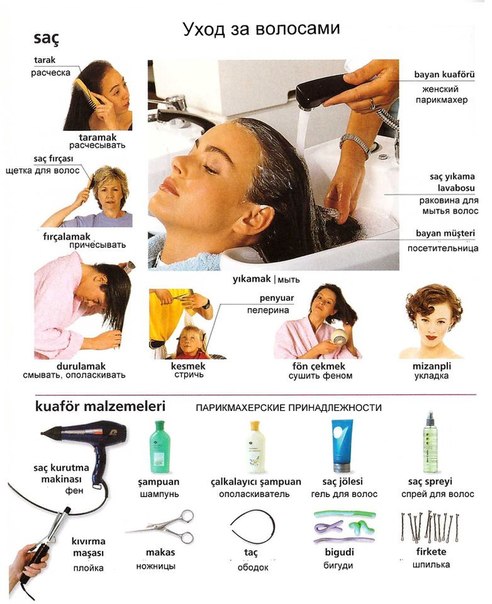
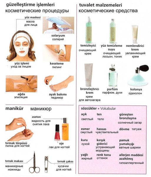
On the subject of beauty and health. Turkish and Russian Language in Pictures.


An abscess is a collection of pus collected in a cavity formed by the
tissue on the basis of an infectious process (usually caused by bacteria
or parasites) or other foreign materials (e.g. bullet wounds). It is a
defensive reaction of the tissue to prevent the spread of infectious
materials to the other parts of the body.


A tooth abscess is a sac of pus (infected material) in a tooth or the
gums that results from bacterial infection.
-

The achilles tendon is the big tendon at the back of the ankle. Tissue banks
frequently 'harvest' this tendon from donors and it is freeze-dried and stored for
transplant (eg to the knee).
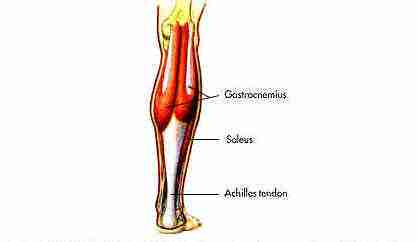
The picture above shows the achilles tendon.
-
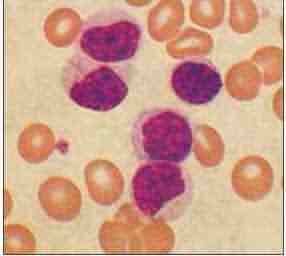
Acute lymphoblastic leukemia, or ALL, is the most common form of childhood leukemia, a
cancer of the white blood cells that invades the bone marrow and crowds out normal healthy
blood cells. This is a type of leukemia that starts from white blood
cells in the bone marrow, the soft inner part of bones. It develops from cells
called lymphocytes, a type of white blood cell central to the immune system, or
from lymphoblasts, an immature type of lymphocyte. The younger the patient is at
diagnosis the better the chances of survival.
Achondroplasia is an inherited disorder of bone growth. It is
one of the group of disorders that are collectively called chondrodystrophies or
osteochondrodysplasias.
The disorder causes a type of dwarfism that is recognized by its characteristic normal
to large-sized head, shortened arms and legs (especially the upper arm and thigh), a
normal-sized trunk, and waddling gait. Achondroplasia is the most common type of dwarfism.
Achondroplasia is inherited as an autosomal dominant trait. However, the majority of
cases, approximately 80%, appear as spontaneous mutations. If one parent has
achondroplasia, the infant has a 50% chance of inheriting the disorder.
If both parents have the condition, the infant's chances of being
affected increase to 75%

-
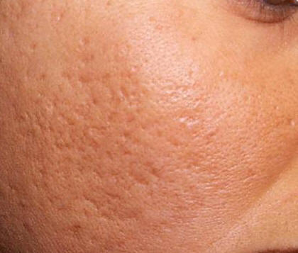
In some people acne can lead to sebaceous cysts; these are sacs just beneath
the skin that are filled with an oily, white, semisolid material called sebum.
You can see in the picture below the swelling and inflammation caused by the
pocket of sebum forming under the skin.

-
The picture to the left is before laser Microdermabrasion.
The picture to the right is after Microdermabrasion

-
Adenoma sebaceum are reddish nodules - angiofibromas - that develop in childhood,
especially on the cheeks and the nasolabial folds in tuberous sclerosis.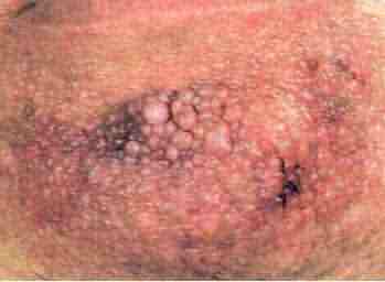
.
Here is a picture of the reddish nodules called Adenoma Sebaceum
-
-
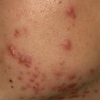
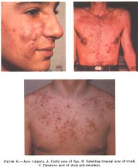
Acne Vulgaris is an inflammatory disease of the skin, caused by
changes in the skin structures consisting of a hair
follicle and its associated sebaceous gland
Acne Vulgaris is common in puberty as a result of an abnormal response
to hormones. Acne Vulgaris usually disappears or decreases as we age.
The age in which is decreases if different in every person.
You can see the inflammation cause by the acne. It presents it self as red
raised areas or spots on the skin.
-
-
Alba Pityriasis is a common skin disease mark by round or oval, finely
scaling patches without skin color (pigment), usually on the cheeks. The itching sores are
easy to spot and occur mostly in children and adolescents. You can see in
the picture below the round patches of skin with out color.
.jpg)
A disease characterized by hypotension, weight loss, anorexia, weakness, and sometimes
a bronze-like melanotic hyperpigmentation of the skin. The picture below
shows the bronze like hyperpigmentation of the skin caused by Addison's disease.

Actinic keratosis is a precancerous skin growth usually caused by sun exposure.
Actinic keratosis occurs most commonly in fair skin, especially
in the elderly and in young individuals with light complexions. The growths occur in
sun-exposed skin areas. The growths begin as flat scaly areas that later develop a hard
wart-like surface.
They are classified as precancerous growths. If left untreated, approximately 10% of
actinic keratosis develop into squamous cell carcinoma.
The picture above shows Actinic Keratosis growths.
-
Adenocarcinoma
is the name of a broad category of cancers. This type of cancer occurs in
cells that line organs such as the colon, lung, and breast
-
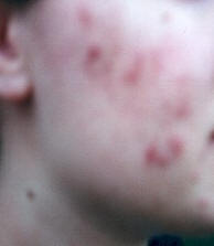
Acne usually
effects people between the ages of 12 and 21. Therefore, it is important
to look for an underlying cause of acne that occurs for the first time
in adulthood when acne that appears after 25 to 30 years of age occurs
for one of these reasons:
Recurrence of acne that cleared up
after adolescence
Flare-up of acne after a period of
relative quiet for example, during pregnancy
Occurs for the first time in a person
who had never previously had acne.
You can see the inflammation
caused by the infection in the skin. It forms raised puss filled bumps on the
skin.
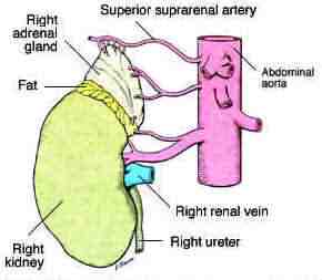
Adrenal glands, which are also called suprarenal glands, are small,
triangular glands located on top of both kidneys. An adrenal gland is made
of two parts: the outer region is called the adrenal cortex and the inner
region is called the adrenal medulla
-
One of the symptoms of AIDS is a rash on the skin. This rash can be a primary symptom
of AIDS or it can result as a side effect of a medication used for AIDS.
-
Agranulocytes are white blood cells that have no distinct granules in their cytoplasm.
-
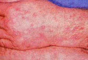
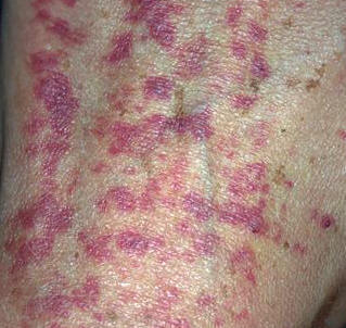
Red, bumpy, scaly, itchy, swollen skin-any of these symptoms can signify
an allergic skin condition. These skin problems are often caused by an
immune system reaction, signifying an allergy. You can see the red inflamed
areas of the skin in the picture below.

Amniocentesis is a diagnostic procedure performed by inserting a
hollow needle through the abdominal wall into the uterus and withdrawing a small amount of
fluid from the sac surrounding the fetus.
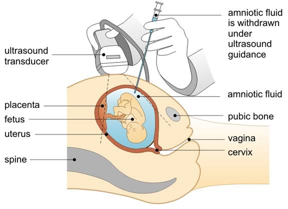
Above is a picture showing the process of an Amniocentesis.
-
Allergic purpura is an allergic disorder. The joints (usually knees, ankles, hips,
wrists, and elbows), the skin of the legs, thighs, and abdomen, the gastrointestinal
tract, and the kidneys are involved. Allergic purpura can affect both sexes, all ages, but
is more common in boys (2 to 8 years).
Purpura is a bleeding disorder that occurs when capillaries rupture, allowing
small amounts of blood to accumulate in the surrounding tissues. In AP, this occurs
because the capillaries are blocked by protein complexes formed during an abnormal immune
reaction. The skin is the most obvious site of reaction, but the joints, gastrointestinal
tract, and kidneys are also often affected.
-
nbsp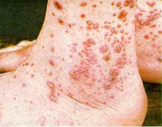
Hypersensitivity to a drug or foreign agent leads to a skin disorder with
inflammation and damage to blood vessels of the skin.
-
The picture below shows the difference
between ambyompia and normal vision.
-
Alopecia areata is the name for a condition in
which round patches of hair loss appear suddenly. The hair-growing tissue is attacked by
the patient's own immune cells for unknown reasons. There are three stages: first, there
is sudden hair loss, then the patches of hair loss enlarge, and last, new hair grows back.
This process takes months, sometimes more than a year, but rarely does the hair never grow
back.
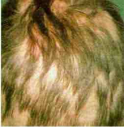
The picture above shows the round patches of hair loss associated with
Alopecia Areata.
-
Total loss of hair of the scalp either within a very short period of time or from
progression of localized alopecia, especially alopecia areata
Patients with alopecia areata lose hair on
their scalp in smooth round patches typically causing bald spots about an inch (2cm)
across. Sometimes the patches are larger, and the condition can include the loss of all
hair on the head (alopecia totalis), and even the loss of hair on the head (alopecia
totalis), and even the loss of all hair on the body (alopecia universalis).
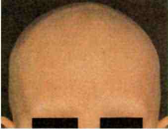
The picture above show total hair loss from Alopecia Totalis
-
Alpha-1 Antitrypsin deficiency is an inherited disorder that may cause lung or liver
disease. Normally, the protein alpha-1 antitrysin, is released into the bloodstream and
travels to the lung where it protects the lungs from the destructive actions of common
illnesses and exposures, particularly tobacco smoke. People with a deficiency of this
protective protein often suffer from progressive lung damage known as emphysema. Unlike
the common form of emphysema seen in otherwise healthy individuals who have smoked for
many years, this alpha antitrypsin deficiency form of emphysema may occur at an unusually
young age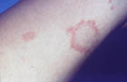
-
The alveoli are the final branching of the respiratory
tree and act as the primary gas exchange units of the lung. The gas-blood
barrier between the alveolar space and the pulmonary capillaries is
extremely thin, allowing for rapid gas exchange. To reach the blood, oxygen
must diffuse through the alveolar epithelium, a thin interstitial space, and
the capillary endothelium; CO2 follows the reverse course to reach the
alveoli
Picture of Alveoli ruptured as a result of emphysema
-
Amyloidosis
Picture
-
Anal cancer, an uncommon cancer, is a disease in which cancer (malignant) cells are
found in the anus. The anus is the opening at the end of the rectum (the end part of the
large intestine) through which body waste passes. It is believe that one of
the main causes of anal cancer is a sexually transmitted disease called HPV and
also anal sex.

-
Google's advertising will not let us keep this picture. Here is a link
where you can find a good
Anal Fistulas Picture
and more
information.
-
-
Angioedema is the development of large welts below the surface of the skin, especially
around the eyes and lips. The welts may also affect the hands, feet, and throat. The
condition can be associated with allergies and histamine release.

-

Angular Stomatitis is an inflammation at the corners of the mouth. It causes
a cracking of the skin. Since it is in the corner of the mouth it makes speaking
and eating uncomfortable.

-
The anterior chamber is the fluid-filled space inside the eye between the
iris and the cornea's innermost surface.
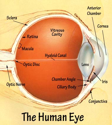
The Anterior Chamber is referenced in the upper right hand corner of the
picture. the anterior chamber is the space between
the cornea anteriorly and the iris/pupil posteriorly, filled with a watery fluid
(aqueous humor) and communicating through the pupil with the posterior chamber.
-
Ankylosing spondylitis
(AS) is a chronic inflammatory form of arthritis that affects the spinal
joints. The hallmark feature of AS is the involvement of the joints at the
base of the spine where the spine joins the pelvis - the sacroiliac
joints.
The disease course is highly variable, and while some
individuals have episodes of transient back pain only, others have more chronic severe
back pain that leads to differing degrees of spinal stiffness over time. In almost all
cases the disease is characterized by acute painful episodes and remissions (periods where
the problem settles). 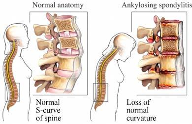
-
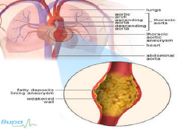
An aortic aneurysm is the dilatation (widening or bulge) of a portion of the aorta,
usually at a weak spot in the aortic wall. The aorta is the largest artery in the body. It
carries all the blood that is pumped out of the " WIDTH="125" HEIGHT="79">
-
Aphthous Ulcers is a canker sore is an open sore in the mouth, which appears as a
painful white or yellow ulcer surrounded by a bright red area. You can see
the ulcer in the roof of the mouth in the picture.

-
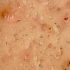
Anthrax is an acute infectious disease caused by the spore-forming bacterium Bacillus
anthracis. Anthrax most commonly occurs in wild and domestic lower vertebrates
(cattle, sheep, goats, camels, antelopes, and other herbivores), but it can also occur in
humans when they are exposed to infected animals or to tissue from infected animals or
when anthrax spores are used as a bioterrorist weapon.

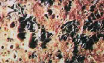
Pictured above is the beginning stages of anthrax.
-